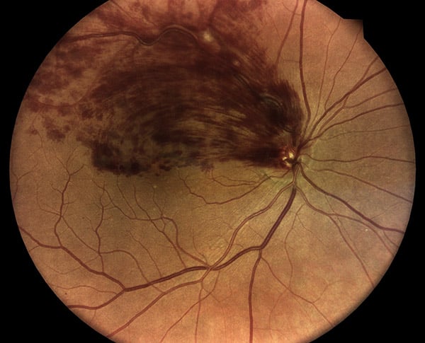
Retinal Vein Occlusions

The retina relies on a network of blood vessels that nourishes its inner layer. Small arteries branch out from the optic nerve into the retina, and the blood they carry is then drained back to the optic nerve and out of the eye through a similar network of veins. Sometimes, one of these draining veins can become blocked by a minute blood clot, often forming at a point where the vein is kinked or bent. When this occurs, the backup of pressure can result in damage to the retina and loss of vision. This usually occurs as a painless spontaneous event which may result in blurred or darkened vision. If the main retinal vein is blocked where it leaves the eye at the optic nerve, it is called a central retinal vein occlusion. If the blockage occurs at a smaller branch, it is known as a branch retinal vein occlusion. These blockages can be partial, so that blood can still flow slowly through the draining vein, or they can be completed, in which case more severe damage occurs to the retina. Depending upon the size and location of the blocked vein, and whether the blockage is complete or not, the occlusion can have very severe consequences to vision, or, on occasion, be completely asymptomatic. If the occlusion is significant, it will cause bleeding and swelling within the retina. In very severe cases, the capillaries in the involved portion of the retina will completely disappear, leaving it starved of blood and oxygen. This can lead to more bleeding within the eye and, in the most serious cases, a very severe form of glaucoma known as neovascular glaucoma. This is where the drain of the eye becomes completely clogged by an overgrowth of abnormal blood vessels and the pressure within the eye rises to very high levels. Untreated neovascular glaucoma will inevitably lead to total blindness within days or weeks.
People at risk for these occlusions are older individuals with a history of high blood pressure, high cholesterol, and diabetes, Open-angle glaucoma, the more common form of the disease, is also a risk factor, particularly for central retinal vein occlusion. Younger middle-aged people can also occasionally be diagnosed with the problem. Very rarely, blood clotting disorders or other blood abnormalities can be associated with retinal vein occlusions, but these are almost never found unless there are other signs and symptoms of clotting problems or multiple recurrent vein occlusions.
There is no perfect treatment for retinal vein occlusions. A mild blood thinner such as low dose aspirin may be recommended in order to help dissolve the blocking blood clot. Many times a three or four-month period of observation may be best, as in a significant portion of patients the problem improves on its own. Probably the best predictor of the possibility of improvement is the initial vision. People who start out with severe occlusion and poor vision do not tend to get better on their own. When treatment is necessary, certain medications may be injected directly into the eye in a procedure called intravitreal injection. This is a quick procedure that is performed in the office and has few side effects. Many patients initially find the idea of these injections quite disturbing, which is a natural reaction. The reality, however, is that the procedure is usually very well tolerated and most patients who undergo it are surprised and relieved at how relatively painless and easy it is. These medications are often effective in improving vision by reducing retinal swelling and bleeding, but they may need to be repeated periodically. Laser treatments may also be recommended, which are also quick in-office procedures. Less commonly, surgery in an operating room may be necessary, especially if severe bleeding or neovascular glaucoma develops. As of yet, there has been no completely safe and reliable way of directly unblocking the occluded vein. Some very delicate and sophisticated surgical methods have been attempted with varying success but are rarely recommended. The visual outcome after retinal vein occlusion, even with appropriate treatment, can be quite variable. Some people have a return of vision to almost normal where others may be left with severe loss. It is important to remember that treatment can be a long process requiring multiple followup visits and occasionally repeated procedures before the disease finally becomes quiet.

